If watching The Pitt is giving you a renewed interest in the human body in all of its gory glory, there’s a new tool that will help satisfy your curiosity. An international team of scientists developed an open-access 3D portal where users can explore human organs in detail. With the Human Organ Atlas (HOA), users can travel through the brain, heart, lungs, kidneys, and liver and better understand our anatomy and the diseases that threaten it. The platform is detailed in a paper published today in the journal Science Advances.
Building on an initial release in 2023, the HOA is now expanded and can be used on a directly standard web browser, without specialised software—or the shrinking technology from Fantastic Voyage. It is powered by an advanced imaging method called Hierarchical Phase-Contrast Tomography (HiP-CT). HiP-CT uses the European Synchrotron’s Extremely Brilliant Source, which is up to 100 billion times brighter than conventional hospital CT scanners. This helped the team scan entire intact human organs without destroying them and then zoom in down to less than one micron, or 50 times thinner than a human hair.
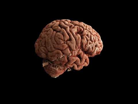
The Human Organ Atlas
According to the team, this technique bridges a 100-year-old gap in medicine between radiology and the study of biological tissue. The project brought together researchers, engineers, clinicians, and infrastructure specialists from nine institutions across Europe and the United States within a consortium called the Human Organ Atlas Hub. It was initially developed during the COVID-19 pandemic, and has already made an impact. It helped scientists detect previously unseen microscopic vascular injury in the lungs of patients who died from COVID-19. Additionally, the advanced imaging is helping cardiologists better understand a normal vs abnormal human heart and being used to probe the step-by-step development of gynecological disorders.
The Human Organ Atlas currently provides access to:
- 56 organs (307 full 3D datasets from 25 donors)
- 11 organ types, including brain, heart, lung, kidney, liver, colon, spleen, placenta, uterus, prostate, and testis
- Multiscale scans, from whole-organ views down to near-cellular resolution
“From the beginning, we wanted these data to be accessible to everyone and build an open, shared scientific infrastructure at a global scale,” Paul Tafforeau, a team member and European Synchrotron (ESRF) beamline scientist, said in a statement. “This is a resource for researchers, doctors, educators—but also for anyone curious about how the human body is built.”

The team also expects that HOA will become a resource for artificial intelligence. Its large high-quality 3D datasets could help train advanced medical AI systems for disease detection and super-resolution analysis.
Importantly, it also offers new opportunities for medical education and public engagement with science.
“Students can explore organs in 3D, scroll through anatomical sections, and zoom into internal tissue detail,” added Alexandre Bellier, an associate professor in anatomy at Grenoble Alpes University Hospital in France. “It creates an immersive exploratory alternative to classic anatomy diagrams, helping learners build a clearer spatial understanding of complex structures. For both teachers and students, it fundamentally shifts anatomy learning from static description to guided, interactive discovery.”
Over the next several years, the team plans to expand the HOA, by adding more organs, samples, and data.

2025 PopSci Best of What’s New